The following scans were performed on an ultrasound club member and uploaded with permission.
Cardiac
This is an apical 4-chamber view of the heart with color doppler. This scan was performed with a cardiac probe at the location of the apical impulse. Note that the probe marker is inverted for this cardiac scan.In the parasternal long-axis view, the mitral and aortic valves are visualized.
FAST scan – Right kidney
This is a view of the right hepatorenal interface, also known as Morrison’s pouch. The focused assessment with sonography for trauma (FAST) scan aims to identify free fluid in the pericardial space or abdomen. The subxiphoid scan can detect a pericardial effusion. The hepatorenal scan can detect fluid in Morrison’s pouch. The splenorenal scan assesses fluid at the splenorenal interface. The pelvic scan aims to assess for free fluid in the rectouterine or rectovesicular pouch (Douglas pouch). Causes of free fluid in the abdomen include trauma and ascites.
Aortic scan – bifurcation
The aortic scan may be used to detect an abdominal aortic aneurysm (AAA). This scan requires visulatization of the aorta starting from the external landmark of the xiphoid process to the aortic bifurcation into the common iliac vessels. A vessel size of ≥ 3cm at any point constitutes AAA.The IVC collapses easily with pressure (shown on image left, anatomic right). The aorta remains round with pressure (shown on image right, anatomic left). The vessels lie anterior to the spine, which casts a shadow posteriorly.
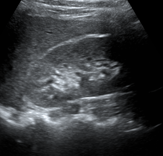
Gallbladder
The gallbladder is visualized in this scan at the center of the image. In addition, the portal vein is the circular struction with a hyperechoic wall left of the gallbladder in this scan. The inferior vena cava is the cylindrical structure right of the liver.
Liver – Hepatic veins
This scan shows the confluence of Right, Middle, and Left hepatic veins, entering the Inferior Vena Cava, which is also shown entering the Right Atrium
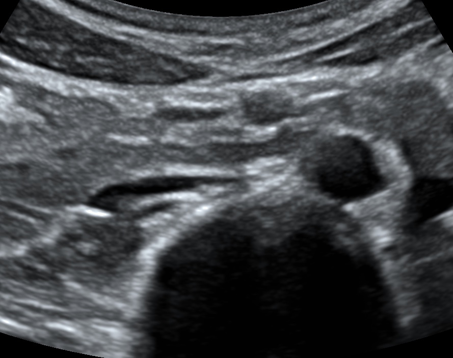
Pancreas
This scan shows the pancreas at the center of the image with the splenic vein coming into view posteriorly.
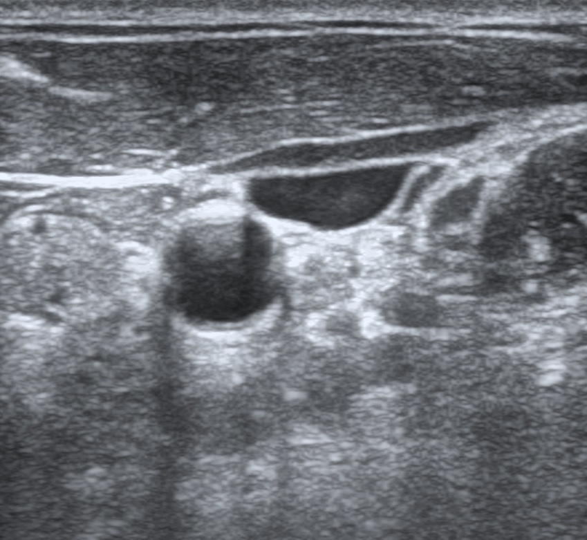
Thyroid
This scan shows the right lobe of the thyroid. The thyroid isthmus and a hyperechoic cartilaginous ring of the trachea is seen medial and anterior to the right thyroid lobe. The common carotid artery is seen pulsating medial to the right thyroid lobe.This is a scan of the left carotid sheath. The sternocleidomastoid is overlying the internal jugular vein, which is collapsable. The common carotid artery is round and does not collapse with compression. The vagus nerve is medial to the carotid and posterior to the IJ.
Aortic arch
This is a suprasternal color ultrasound of the aortic arch, and also showing flow in the left common carotid artery.